Vibrationnal Spectroscopy
Presentation
The Vibrational Spectroscopy Division hosted at LASIRE is one of the 10 divisions of the Advanced Characterization Platform of the Institut Michel-Eugène Chevreul (CNRS FR2638).
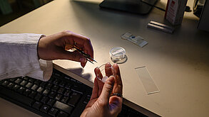
The division brings together Raman micro-spectrometry and Fourier Transform Infrared (FTIR) spectroscopy at LASIRE. It is equipped with nine FTIR spectrometers and Raman microspectrometers, as well as an infrared microspectrometer for the analysis of micro-samples. Thanks to the variety of accessories available, the division allows the analysis of diverse samples in different physical forms (liquid, solid, gas…) and under varied experimental conditions (temperature effects, under irradiation…).
The division provides services or collaborations for sample characterization, identification, molecular reactivity analysis, etc. Personal or group training in vibrational spectroscopy techniques can also be offered.
Expertise
The areas of application are varied:
- Materials: Characterization of chemical composition, defects, pollution, or the origin of aging in materials (glass, textiles…)
- Environment: Characterization of pollutants or contaminants, detection of product modifications
- Microelectronics: Characterization of materials, quantification of stress during operation
- Pharmacy and Medicine: Characterization of compounds, including the distribution of active ingredients, interaction of molecules with tissues or cells
- Geology: Analysis of minerals, fossils, identification of gemstones and pigments on art objects
- Food Industry
For any analysis request, please send an email to the following address: pole-spectroscopie-vibrationnelle_at_univ-lille.fr
Location / Address:
LASIRE – Building C8 – Cité scientifique
59655 Villeneuve d’Ascq
Staff:
Isabelle De Waele (Scientific Head of the cluster)
Myriam Moreau (Technical Head of the cluster)
Main Equipment:
The instrumental cluster is equipped with 4 Raman microspectrometers, 5 Fourier Transform Infrared (FTIR) spectrometers, and 1 FT-Raman spectrometer.
Presentation
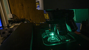
Raman scattering enables the molecular analysis of solid, liquid, or gaseous samples.
Generally non-destructive, this technique allows the identification and localization, at the microscopic scale, of the different constituents of a heterogeneous sample of macroscopic size.
Raman spectroscopy can be used to study molecular organization, crystallinity, stress, electronic properties, etc. Its complementarity with IR lies in the better detection of vibrations originating from weakly polar groups, for example C=C, S–S, etc.
Technical Manager: Myriam Moreau (Myriam.Moreau_at_univ-lille.fr)
Equipment and Technical Description

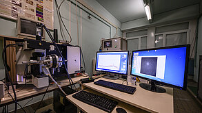
HR Visible Spectrometer
| Components of the Spectrometer | Features |
|---|---|
| Spectrograph | 800 mm focal length spectrograph 3 gratings : 1800 t/mm - 600 t/mm - 300 t/mm |
| Laser | 632.8 nm, 473 nm, 532 nm |
| Objectives | ×10, ×50, ×100, 50X LWD, ×100 LWD, 90° prism for liquid observation |
| Available Accessories | Motorized XY stage – Nano-positioning stage Linkam stage: temperature control from –180 °C to 600 °C Double scanner: illuminates a surface with the laser instead of a point Half-wave plate and analyzers |
HR Evolution Spectrometer
| Components of the Spectrometer | Features |
|---|---|
| Spectrograph | 800 mm focal length spectrograph 3 gratings : 1800 t/mm - 600 t/mm - 300 t/mm |
| Laser | 632.8 nm – 515 nm – 785 nm diode laser |
| Objectives | ×10, ×50, ×100, 50X LWD, ×100 LWD, 90° prism for liquid observation |
| Available Accessories | Motorized XYZ stage Linkam stage: temperature control from –180 °C to 600 °C Half-wave plate and analyzers |
HR UV Spectrometer
| Components of the Spectrometer | Features |
|---|---|
| Spectrogaph | 800mm focal length spectrograph 2 grattings: 2400 t/mm - 600 t/mm |
| Laser | 244nm - 325 nm |
| Objectives | 40X – 15X – 80X |
| Available Accessories | Motorized XY stage Linkam stage: temperature control from –180 °C to 600 °C |
LabRam Spectrometer
| Components of the Spectrometer | Features |
|---|---|
| Spectrograph | 300 mm focal length spectrograph 2 gratings : 1800 t/mm - 300 t/mm |
| Laser | 632.8 nm |
| Objectives | ×10, ×50, ×100, 50X LWD, ×100 LWD, 90° prism for liquid observation |
| Available Accessories | Motorized XY stage Linkam stage: temperature control from –180 °C to 600 °C Half-wave plate and analyzers |
Presentation
The center has several FTIR spectrometers covering the mid-infrared (MIR) range (4,000 to 450 cm-1) and the near-infrared (NIR) range from 1 µm to 2.5 µm (10,000 to 4,000 cm-1), as well as an infrared microspectrometer.
Equipment
The center is equipped with four spectrometers:
- Vertex 70 spectrometer coupled with an infrared microscope (Hyperion 30000) (Bruker) Vertex 70V spectrometer (Bruker)
- Tensor 27 spectrometer (Bruker)
- RFS 100/S FT Raman spectrometer (Bruker)
Accessories
ATR cells, DRIFT, Praying MantisTM reaction chamber (HARRICK), Linkam heating cell (FTIR 600H), etc.
Technical Manager: I. De Waele
IR Microscope Hyperion 3000 coupled with Vertex 70 spectrometer (Bruker)
FTIR Spectrometer Configuration:
| Sources | Mid-infrared Near-infrared |
|---|---|
| Beamsplitters | KBr (Mid-infrared) CaF₂ (Near-infrared) Automatic beamsplitter selection |
| Detectors (FTIR Spectometer) | DTGS [4 000-350 cm¹ ] MCT [12 000-600 cm¹ ] |
| Scan Speed | Rapid Scan module allowing acquisitions up to 56 spectra/s |
Microscope Configuration:
| Objectives | 15X, 36X ATR Imaging Accessory (SIL) equipped with a 20X objective |
|---|---|
| Detectors | Mono element:
Extended Focal Plane Array (FPA) detector:
|
Tensor 27 Spectrometer, Bruker
This device is designed for the mid-infrared range and is equipped with a DTGS detector and an MCT detector.
It features a KBr beamsplitter, allowing spectra to be obtained with a resolution of up to 0.5 cm⁻¹.
It is equipped with a diamond ATR accessory and a module for performing transmission measurements.
FT-Raman Spectrometer RFS 100/S, Bruker
Although dedicated to Raman spectroscopy, this spectrometer is part of the platform due to the technique used (Fourier Transform).
It can operate continuously for several days thanks to the large-capacity dewar used to cool the detector.
Vertex 70V Spectrometer, Bruker
FTIR Spectrometer Configuration:
| Sources | Mid-infrared |
| Beamsplitters | KBr (Mid-infrared) |
| Detectors | DTGS [4,000–350 cm⁻¹] MCT [7,000–600 cm⁻¹] |
| Scan Speed | Rapid Scan module allowing acquisitions up to 56 spectra/s Step Scan module enabling time-resolved acquisitions with a resolution down to a few nanoseconds |